Squamous Cell
Carcinoma.
SCC is a major type of skin cancer.

SCC is a major type of skin cancer.
Squamous Cell carcinoma (SCC) is a cancer of keratinocytes which are the cells found in the thin top layer of the skin called the epidermis.
SCC is the second most common type of non-melanoma, after BCC.
This article refers to “Invasive SCC.” The other type of SCC is Intraepithelial Carcinoma (IEC). The appearance and treatment of invasive .v. IEC is different. The cancerous cells of an invasive SCC have spread into the dermis of the skin, whereas the cancerous cells of an IEC remain in the epidermis (uppermost layer).
Invasive SCC often occurs after the age of 40 and is 3 times more common in men than women.
The areas most commonly affected are the head and neck and other sun-exposed areas of the body. This means an SCC is regularly seen in those with an outdoor occupation or recreational activity. People with fair skin, blue eyes and blonde or red hair have a higher risk of developing an Invasive SCC and smoking can also be a factor.
SCC usually develops in a preexisting Solar Keratosis, and previous skin cancers are strong predictors for subsequent ones.
SCC typically has at least one of these features:
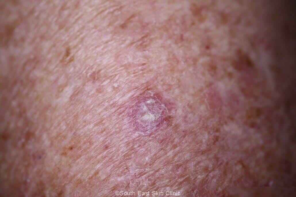
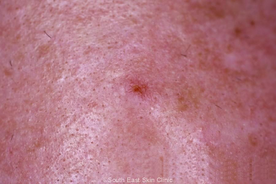
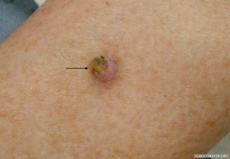
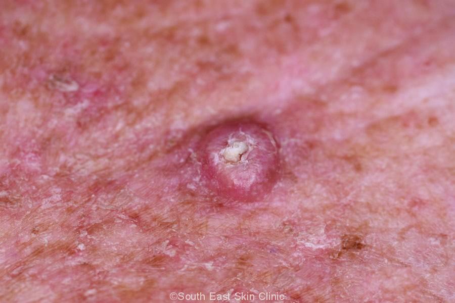
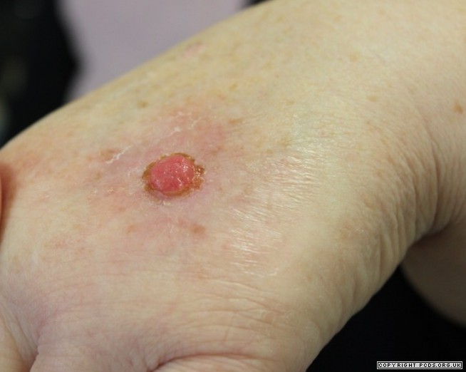
The appearance of an early SCC is similar to that of a thick (Hypertrophic) Solar keratosis. An invasive SCC will typically be elevated and scaly once it has progressed. At this point, the base will be red and feel firm and it can look very similar to a keratoacanthoma.
Occasionally, invasive SCCs are not scaly. Their appearance is similar to that of nodular melanoma. Dermoscopy is very helpful, although a biopsy will be required to make the final diagnosis and determine the need for removal.
Gallery of SCC
Invasive SCC will need to be surgically removed with a margin of clinically normal-looking skin around the lesion. This margin is usually larger than what is required for a BCC. The exact margin chosen is a judgment that takes into consideration the risk factors for aggressive SCC.
It’s helpful to consider the risk factors for SCC recurrence before biopsy and the risk factors from the pathology report.
The risk factors for a more aggressive SCC that are known prior to surgical excision are:
The standard excision margin is around 3-4mm, but that precise figure will depend on clinical features and patient preference.
Following excision with margins, will any further treatment (e.g. surgery) be required? The risk factors in the pathology report for an aggressive SCC are:
The latest guidelines highlight the importance of the depth of an SCC. Depth is measured using the “Breslow thickness,” which is the distance from the top of the skin (almost) to the deepest level of The SCC. The Breslow thickness is also used to stage melanoma, and its use to help stage SCC is a recent development. A Breslow thickness of at least 2mm indicates an SCC that may be more aggressive.
Further treatment (Surgery) will often be required when the above pathological features are identified.
The excision margin required for SCC will tend to be greater than for a BCC
These are some of the key terms that may be used in the SCC Pathology Report.
The degree of differentiation of the cells is an important piece of information.
Atypical keratinocytes
Atpical means that the keratinocytes are not typical eg. the cells look glassy, and their nuclei look abnormal. Some divide by mitosis abnormally.
Keratinocytes extending into the dermis
The Keratinocyte cells do not look typical and they extend into the dermis. The keatinocytes clump together as island that are found in the dermis.
Keratinization
Keratin forms in excess amounts. This accounts of the scale. The keratin may found in cells or outside cells. When keratin is found in large amounts forms a keratin horn (which are also found with keratoacanthoma). A keratin horn is a visible projection of scaly skin.
Differentiation
The cells may appear the same as normal keratinocytes (well differentiated), not quite the same (medium differentiation) or very different (poorly differentiated). Poorly differentiated indicates that the tumour cells are dividing more rapidly and may require additional monitoring or further treatment.
Well differentiated SCC cells may produce quite a lot of keratin ie. scale.
Perineural Invasion
Perineural invasion (PNI) indicates invasion of nerve cells. Perineural invasion is more likely to be found in elderly people, and tends to involve the head and neck. They are found in around 5% of invasive SCC.
Perineural invasion is an important risk factor for aggressive SCC.
PNI involving nerves of greater than 0.1mm has a higher rate of recurrence and metastasis.
Breslow Thickness
Breslow thickness is the distance between the top of the skin and deepest area of the SCC.
The upper layer is fixed and is found at the top of the granular layer of the epidermis – this is very close to the surface of the skin.
A Breslow thickness of >2mm is a risk factor for a more aggressive SCC.
SCC is very common in older Queenslanders with sun-damaged skin.
 ©South East Skin Clinic, All rights reserved
 ©South East Skin Clinic, All rights reservedDermatofibroma