The Common Mole.
How do you spot a harmless one?

How do you spot a harmless one?
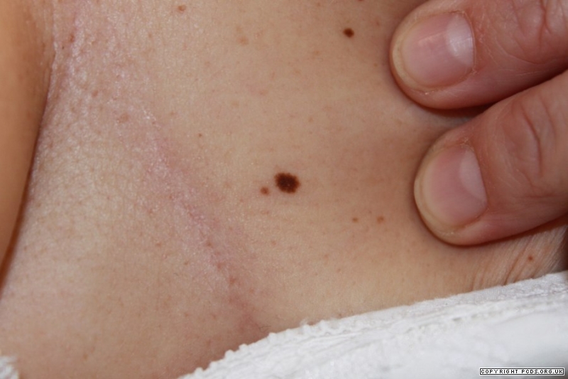
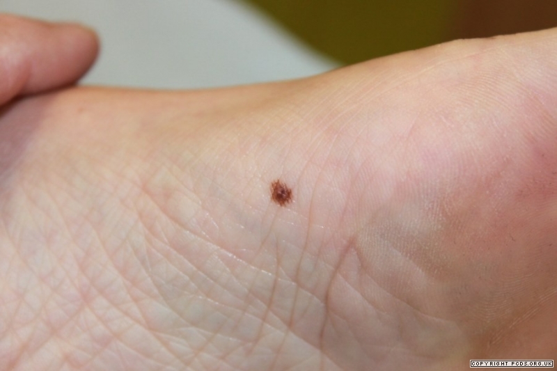
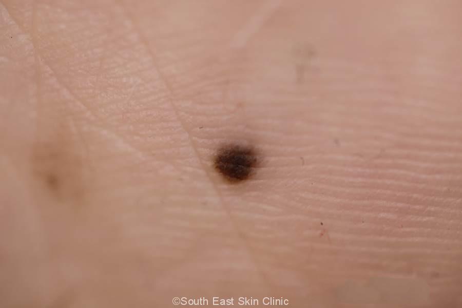
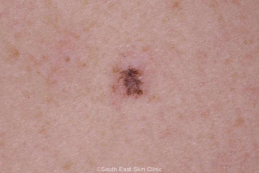
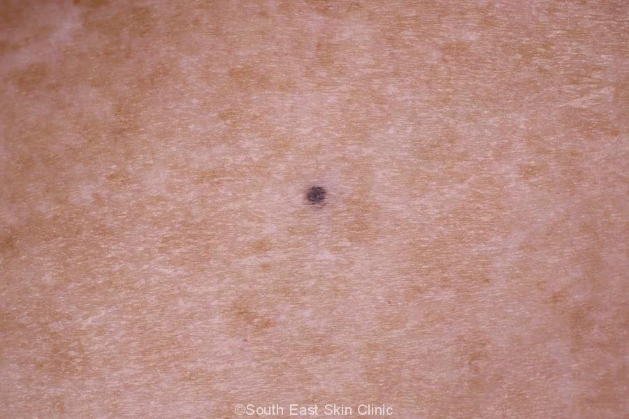
A mole (nevus or naevus) is a benign ‘nest’ of melanocytes, which are cells that produce melanin. Melanin is the pigment that causes skin colour. Looking closely at each mole is a crucial part of a skin check.
Everyone has moles, and they were probably determined before you were born. Typically, moles appear in childhood, but they may also appear throughout your life, particularly during times of hormonal changes. Fair-skinned people tend to have a larger number of moles than darker-skinned people.
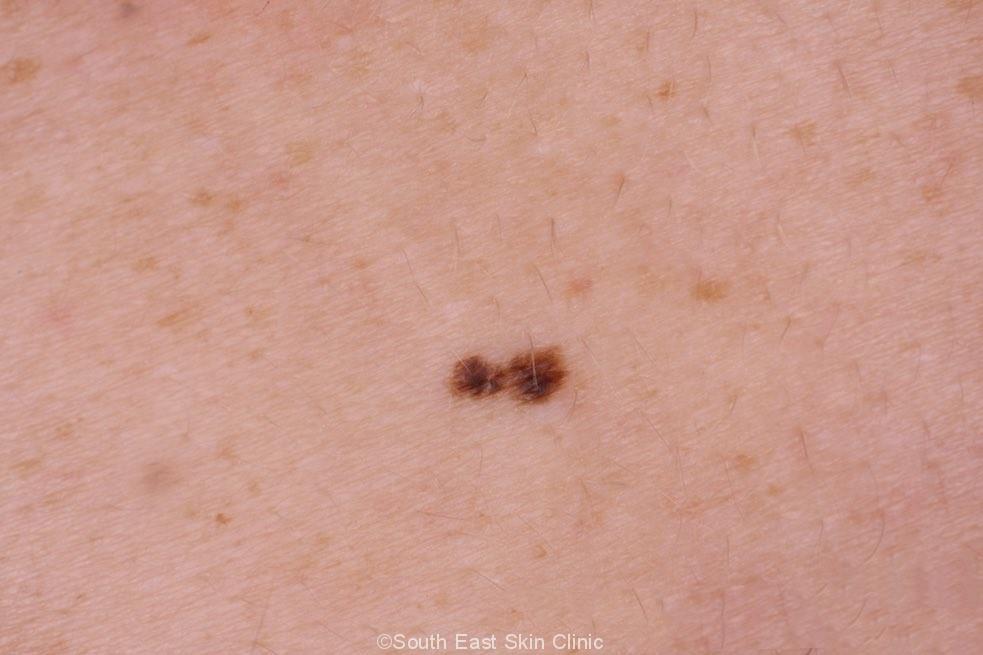
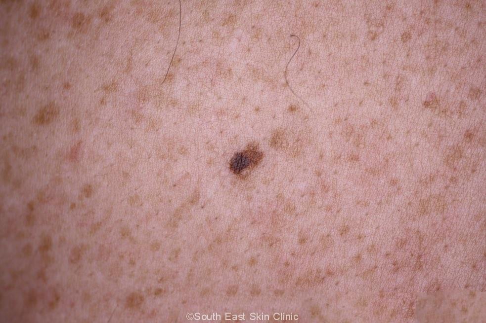
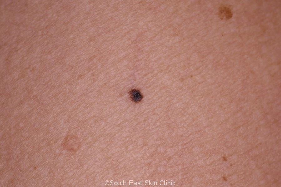
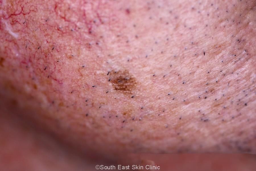
A mole is simply an accumulation of melanocytes, the pigment-forming cells of the epidermis (outer layer of skin). These nests of melanocytes come in a bewildering variety, and the easiest way to classify them is by the position of the nests within the skin. The nests can be superficial, deep, or a mixture of the two and include:
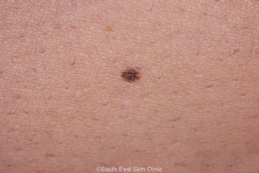
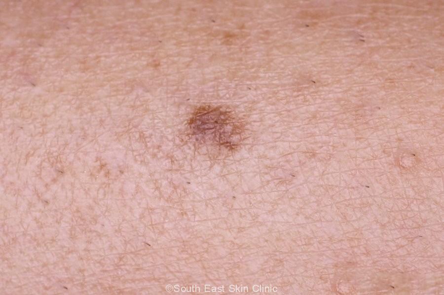
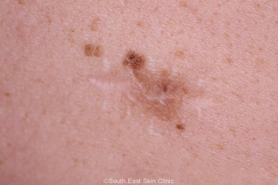
A mole can be identified as Junctional, Dermal or Compound by its dermoscopic appearance and a biopsy would confirm the diagnosis. Ultimately, what matters is that the mole is not melanoma!
There are several different types of moles, including:
What do the different types of Moles look like?
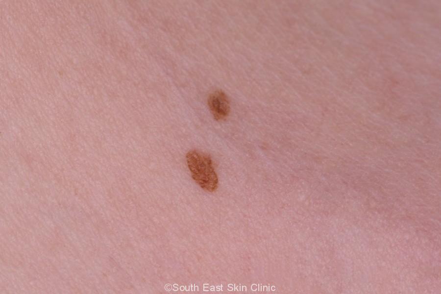
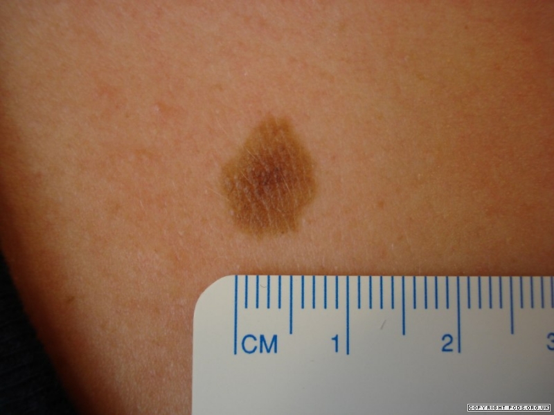
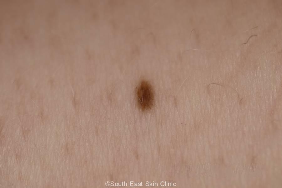
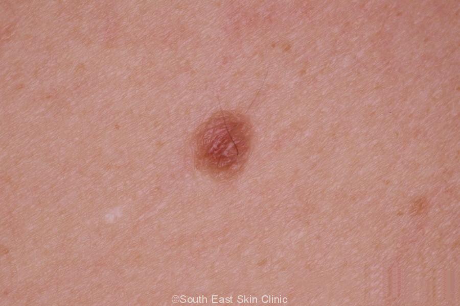
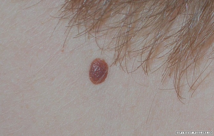
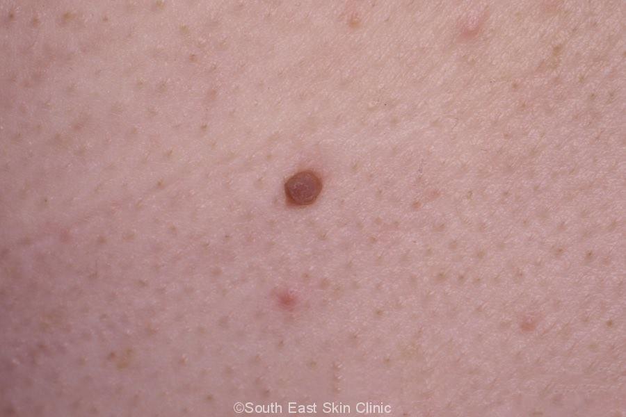
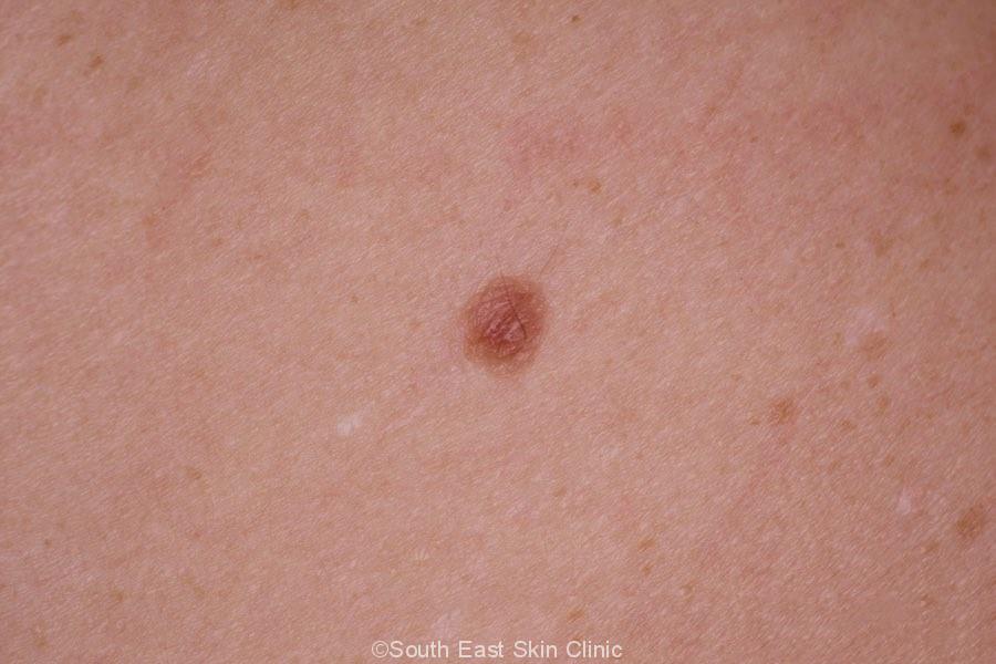
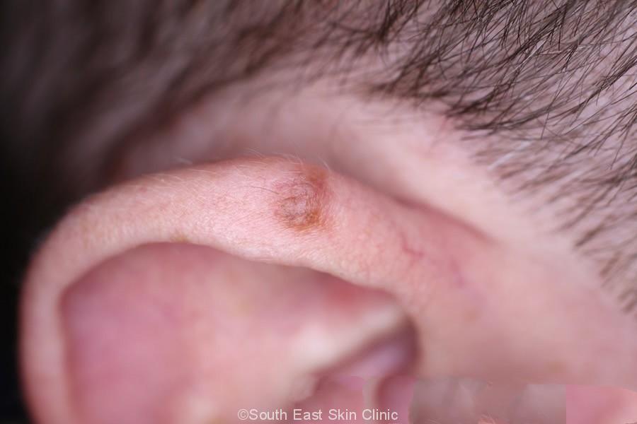
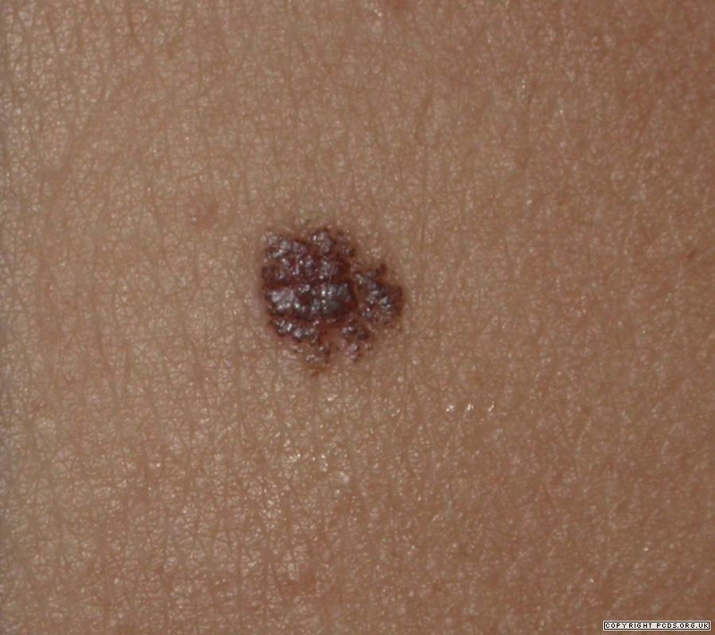
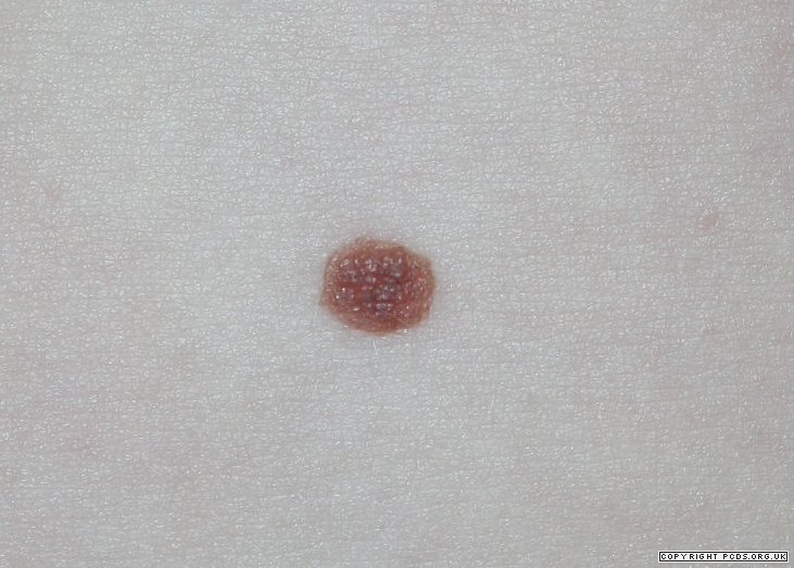
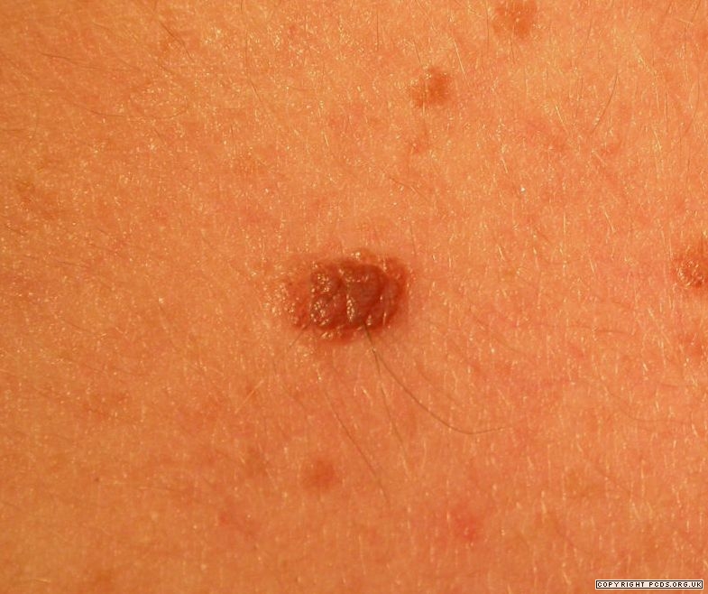
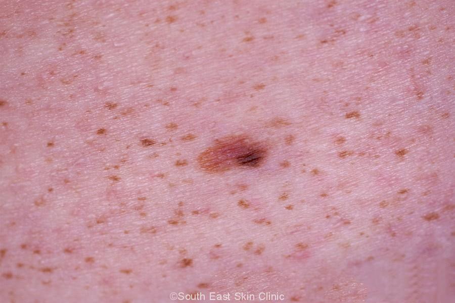
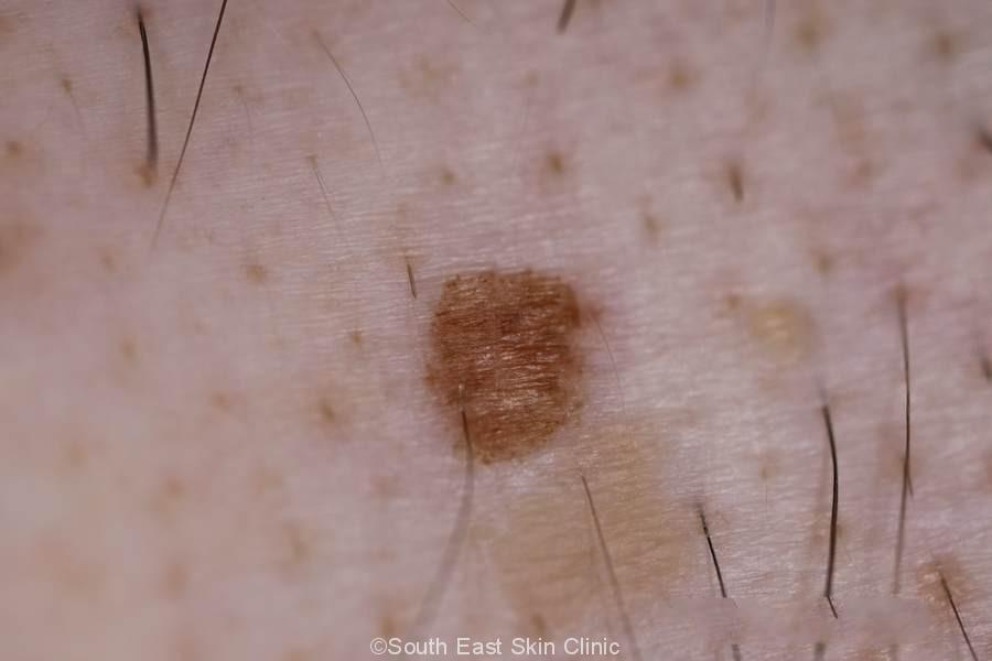
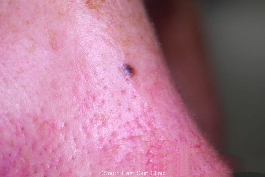
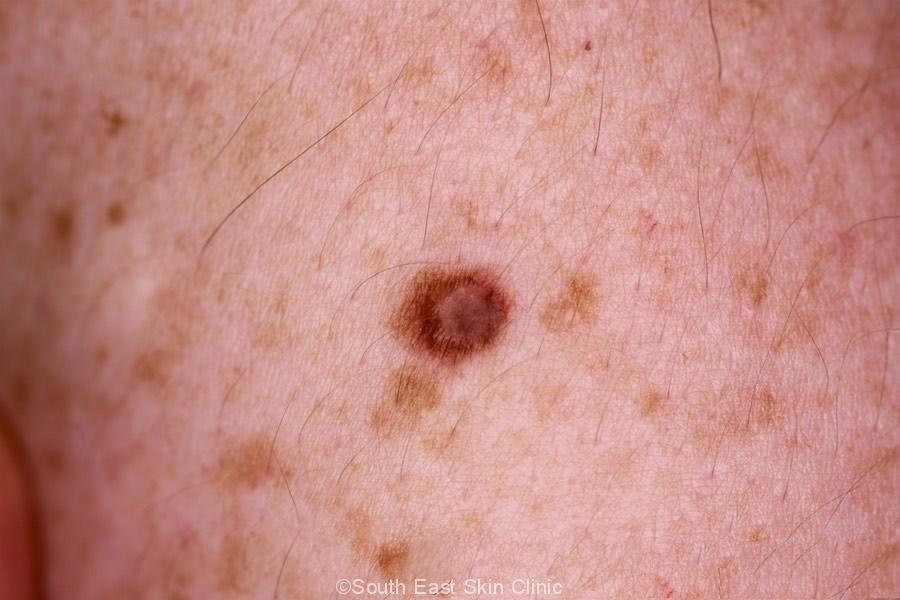
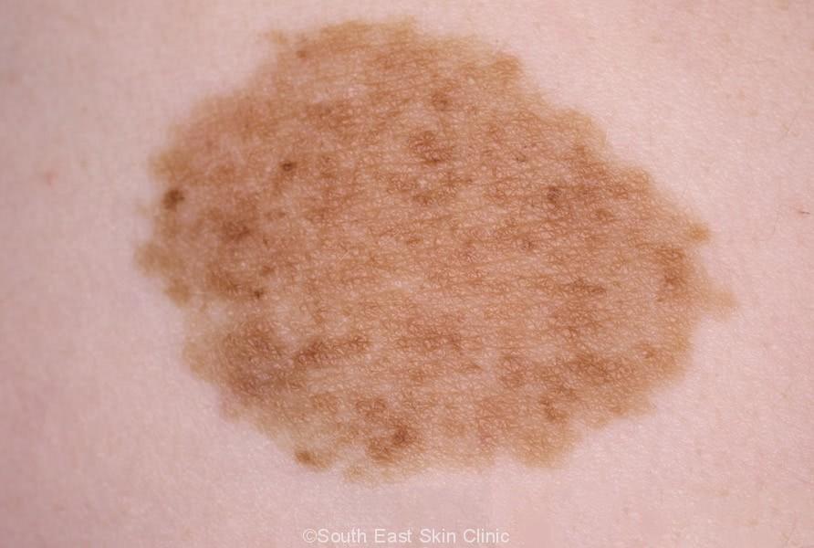
The appearance of a mole depends on whether it is junctional (flat & involving only a thin upper layer of skin), dermal (thick & involving deeper layers of skin) or compound (in-between).
That is why your moles may all appear so different from one another.
Flat Moles (Junctional Nevi)
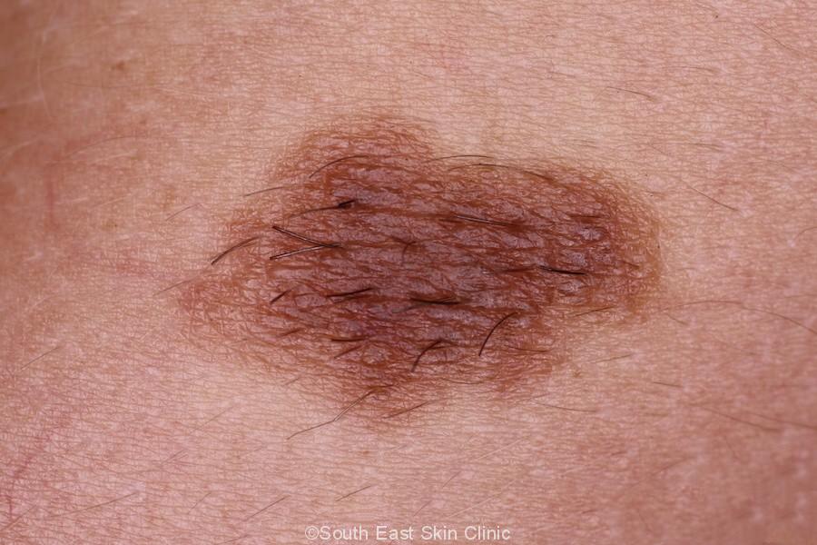
Thick moles (Dermal Nevi)
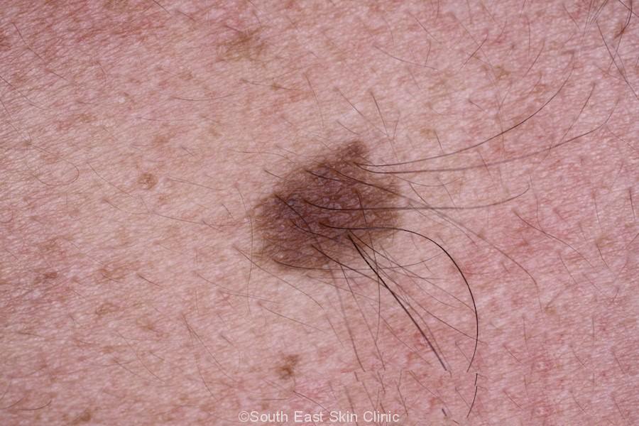
Compound Moles
This type of mole combines the two types mentioned above (Junctional moles and Dermal moles). They usually appear raised and dark and can have features similar to melanoma.
Congenital nevi moles usually appear at birth or in infancy.
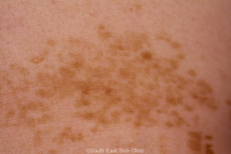
Speckled nevus moles present exactly as their name suggests and recurrent nevis moles which have regrown after an incomplete removal.
Facts about Moles and Melanoma
If everyone has moles, then you might wonder why they matter. While it’s true that most moles are harmless, here are some less benign facts about them:
Melanoma may develop from a mole – although it’s more common for a melanoma to develop spontaneously.
After an excision and biopsy of a mole, you’ll get a Pathology Report detailing the results. A nevus is only removed for either cosmetic concerns or to exclude a melanoma (or another type of skin cancer).
Junctional Nevus
In a junctional nevus, the nests are located along the base of the epidermis.
Compound Nevus
The melanocyte nests are found both in the lower layer of the epidermis (as for a junctional nevus) and also the dermis (as for a dermal nevus) – it’s basically a combination of junctional & dermal.
Dermal Nevus
The nests of melanocytes are found in the dermis ie. in the deeper layer of skin than with a junctional nevus.
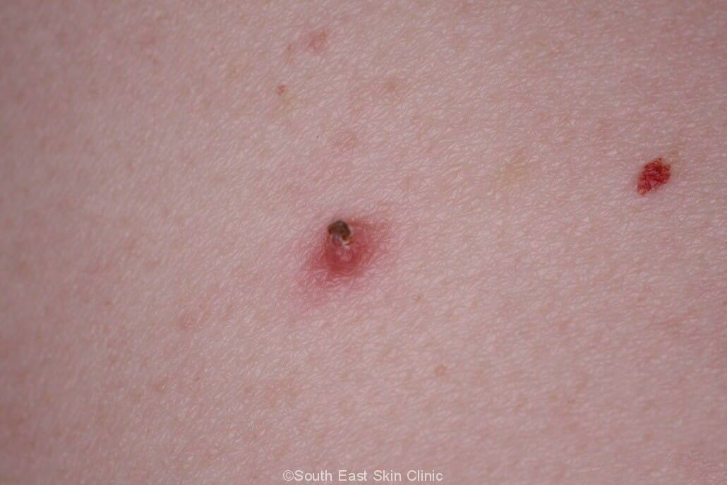
Dysplastic Nevus
Dysplasia is an important term in pathology reports. Dysplastic melanocytes look abnormal in some way.
The cells may have a nucleus that’s abnormal in shape and colour. The nuclei may be angulated, a darker colour (hyperchromasia), or it may nucleoli may be prominent. There may also be bridges between epidermal projections, which are finger-like projections into the epidermis from the layer below, or even some fibrosis (scar-like tissue).
If the pathologist can’t tell a lesion from a melanoma, then it’s called a severely dysplastic nevus.
Moles are the most common of the benign lesions.
 ©South East Skin Clinic, All rights reserved
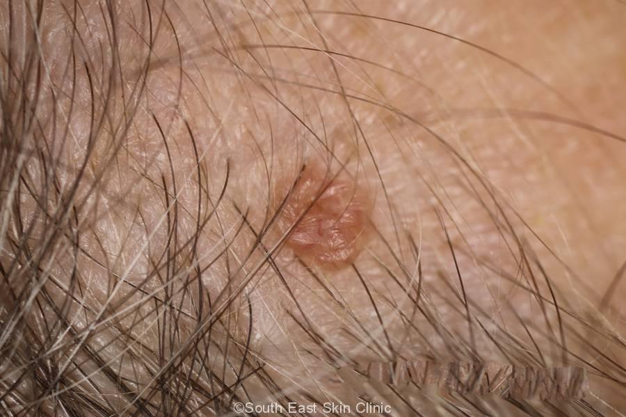
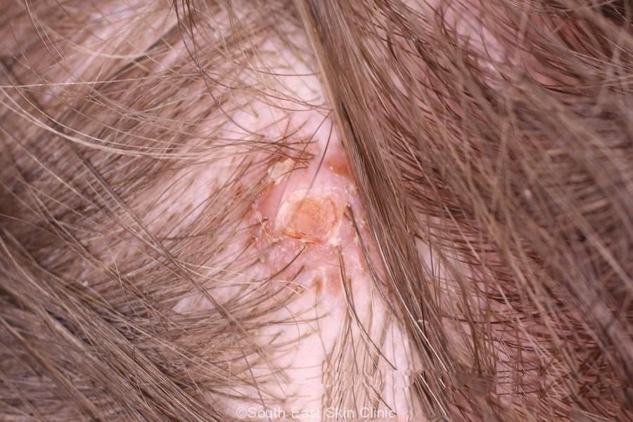
 Folliculitis