Solar
Keratosis.
Sunspots are uiquitous in Australians over the age of 40.

Sunspots are uiquitous in Australians over the age of 40.
Solar Keratosis are known as Sunspots. These rough, scaly lesions found on the backs of hands and forearms or on the face and forehead are very common in Australia!
The p53 gene codes a protein that suppresses the abnormal growth of cells. UVB damages this tumour suppressor gene so that abnormal cells start to appear, manifesting as inflamed, scaly skin called ‘sunspots.’
Solar keratosis is almost to be expected if you were brought up in Australia, have fair skin, and are over the age of 40 to 45.
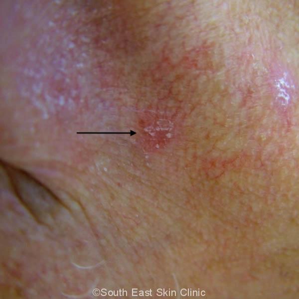
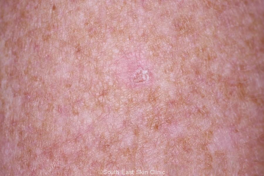
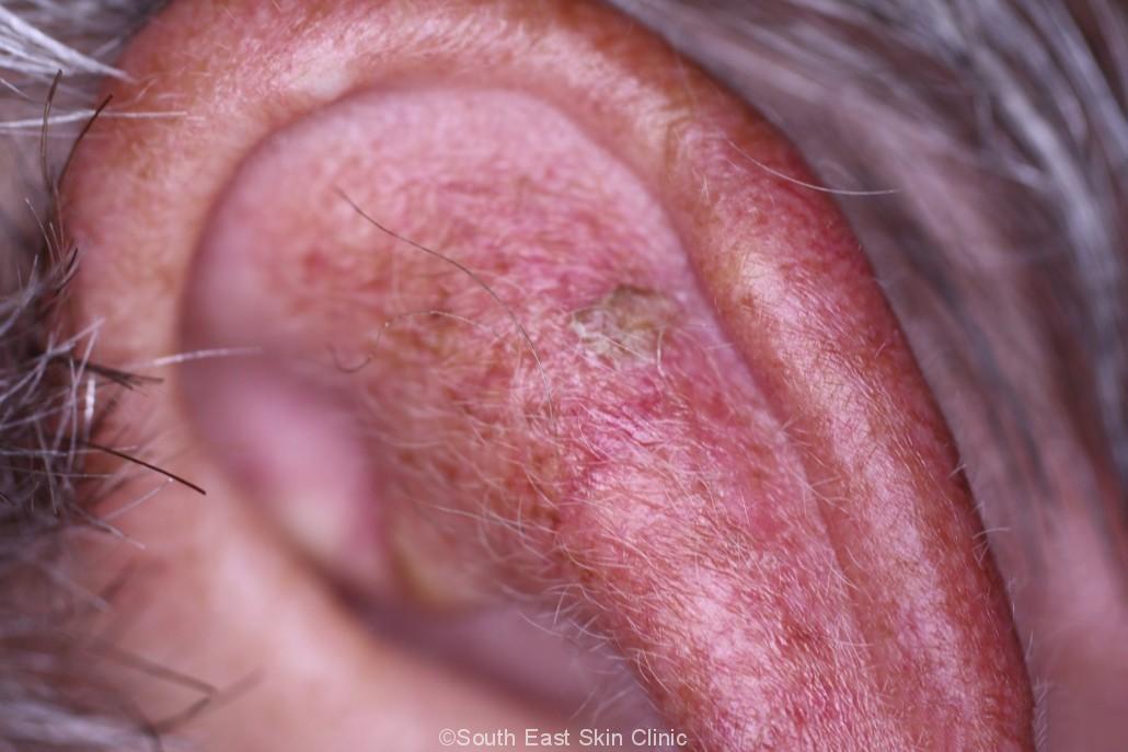
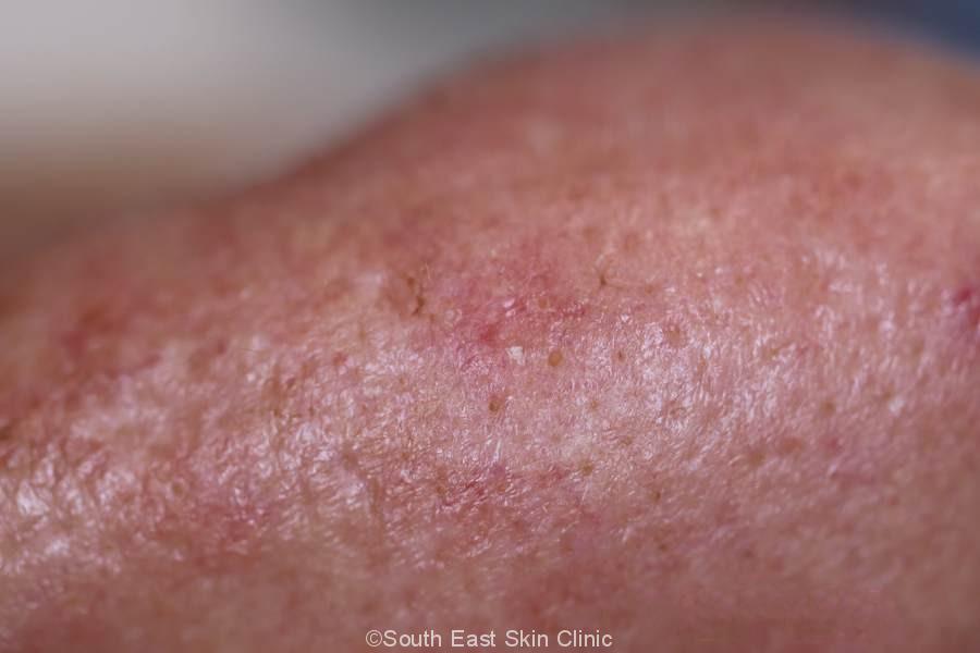
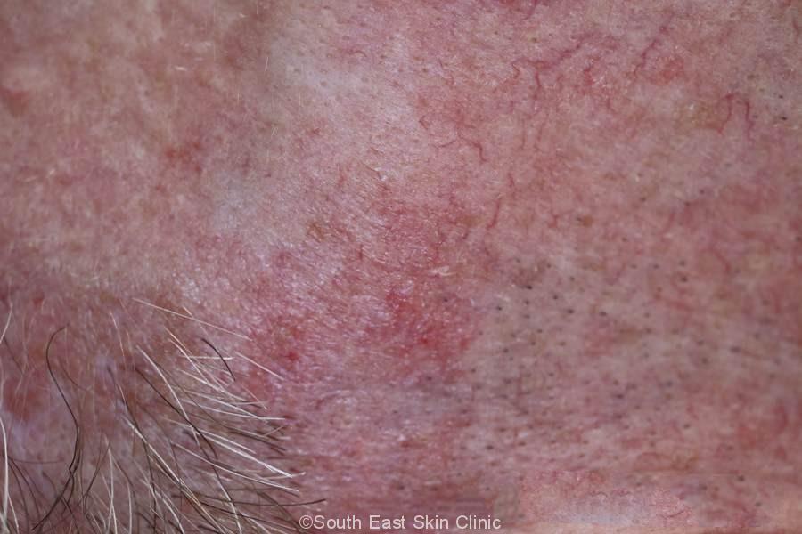
The typical solar keratosis will be a red flat scaly area on the back of the hands, forearms, face or scalp (in men with hair loss). They are often better felt than seen. A sunspot is usually less than 1cm in diameter, although they may be grouped together as an almost continuous area of ‘field change’ that reflects an area of UV-damaged skin.
The appearances can vary greatly. There are different subtypes depending on their clinical appearance:
In order to determine whether the lesion is Solar Keratosis and not skin cancer or some other alternative diagnosis, a dermatoscope may be required to examine the lesion closely. If there is any doubt, a biopsy will be required.
Gallery of Sunspots
Please click on the images for details.
Why do sunspots matter if so many of us get them?
Because of the risk of transition of Solar Keratosis to Squamous cell carcinoma, areas of solar keratosis are generally treated. The precise risk of a specific untreated Solar Keratosis developing into SCC is not known for certain. One figure from a study suggests a 10% chance that a person with 7 to 8 untreated Solar Keratoses will develop one SCC within 10 years. Research suggests that 60 to 80% of SCC arises from solar keratosis.
Some Solar Keratoses may go away on their own without any treatment other than good regular sun protection.
Treatment may be targeted to individual Sunspots or to a whole ‘field’ such as the face, scalp, forearm and/or back of hands. Treatment of a whole field will reduce the risk of solar keratosis appearing in those adjacent areas in the future. When in doubt, it’s generally best to treat a whole field.
Have you seen someone walking around with a crusty red nose, cheeks, forehead and/or temples? Most likely, they are having field treatment for Solar Keratosis.
There are over ten different treatment options available. The most widely used treatments are efudix and cryotherapy.
Many regular attendees of a skin cancer clinic are familiar with Cryotherapy (freezing). The issue with cryotherapy is that it does not treat a whole field. Efudix cream, on the other hand, does treat a field but usually involves several weeks of red and inflamed skin. Aldara and Picato gel are limited by the amount of skin that can be treated at a time. None of these situations are ideal, which is the reason why people often choose PDT. However, PDT is still expensive.
Physical treatments may be used to treat individual Solar Keratosis or whole field:
There are other topical therapies for mild solar keratosis
Regular use of a moisturizer is helpful.
Surgical treatments may be used to treat more isolated lesions:
Most prescriptions for Picato® gel & Aldara® are on a private prescription because PBS (Medicare) requires that the treatment is restricted to the face or scalp and that other standard treatment is ‘inappropriate.’
Solar Keratosis may be difficult to control in some people. The starting point at home is a moisturizer, sun protection and Vitamin B3 500mg twice per day.
Treatment of Solar Keratosis is worth the effort because of fewer sunspots and less skin cancer in future.
| Effectiveness | Treatment Schedule | Side Effects Severity | Duration of Side Effects | Cosmetic Outcome | Price | Treatment Area | |
|---|---|---|---|---|---|---|---|
| Efudix (5FU) | Moderately effective | 2-4 weeks of twice daily application | Marked | 1-2 weeks after stopping treatment | Usually Good | $ | Large areas |
| PDT (Photodynamic Therapy) – clinic | Effective | one or two visits | Moderate but can be painful | 1-2 weeks | Good to Best | $$$$ | Large areas |
| PDT (Photodynamic therapy) – daylight | Effective | one or two visits | Mild to Moderate | 1-2 weeks | Good to Best | $$$ | Particularly good for the face and scalp |
| Aldara | Moderately Effective | 3x overnight applications per week for 4 weeks | Marked | 1-2 weeks after stopping treatment | Usually Good | $$ | Small – Maximum treatment area: 5cm by 5cm |
| Picato Gel | New to the Market – moderately effective compared to placebo | 2 -3 days with once daily applications | Marked | 7-10 days | Should be the same as Efudix or Aldara | $$$ | Small – Maximum treatment area: 5cm by 5cm |
| Solaraze Gel | Generally Not considered very effective | 3 months of twice daily applications | Minimal | Treatment Duration | Neutral | $ | Small to Moderate |
Treatment is usually based on clinical appearance. On occasion, a solar keratosis is surgically removed.
These are some of the key terms that may be used in the Solar Keratosis pathology report.
Atypical Keratinocytes
The keratinocytes are the cells found in the base of the epidermis. Atypical Keratinocytes look abnormal (the cells are large and pink) because of UV damage. This is why some lesions, left untreated, would have gone on to develop IEC & SCC.
The abnormal keratinocytes are the same abnormal keratinocytes found with an IEC – the difference being that the abnormal keratinocytes of an IEC extend throughout the epidermis (“full thickness atypia”). The abnormal keratinocytes of a Solar Keratosis do not extend more than two thirds upwards into the epidermis. In addition, the abnormal keratinocytes do not invade downwards into the dermis (or it’s otherwise an SCC / squamous cell carcinoma).
Dysplasia
Dysplasia indicates that the cells are abnormal – the cells in question being the Keratinocytes. There is a continuum of dysplasia with mild dysplasia, moderate and severe dysplasia on the one hand and actual skin cancer on the other (IEC or SCC).
Abnomal Keratinisation
Keratinocytes produce keratin. Excessive production of keratin leads to a scaly thickened epidermis and this is called abnormal keratinisation. The scale may become thick in which case it will be obvious when you look at the lesion – the scale of a “hypertrophic” solar keratosis can almost be picked off. Very thick lesions are called cutaneous horns and project upwards from the skin as a hard scaly nodule.
Parakeratosis
The keratinocytes normally migrate upwards from the base of the epidermis to the uppermost layer called the stratum corneum – and lose their nuclei in the process. Parakeratosis indicates that the cells have not all lost their nuclei in the uppermost layers of the epidermis.
When you look at the evidence, this is simple. Good Quality Sun Protection will considerably reduce the risk of further Sunspots developing, and of course, reduce the risk of melanoma and other skin cancers. In terms of Sunspots, The seminal Nambour Study² really should be more widely known. This is an old study simply because it would be unethical to repeat.
There were 588 participants in the Nambour study. Each participant lived in & around Maryborough. At the beginning of the study, each participant had between 1 and 30 Sunspots. They were each asked to put on a cream once every morning, with “approximately 1.5 ml to the head and neck and the same amount to each forearm and hand once … and to reapply if necessary during the day.” (Bear in mind that modern sun protection advises 3mls on each of these body areas with very frequent re-application).
Half of the participants used sun protection, 17 sunscreen, and the other half used a sham-cream. They didn’t know which they were using.
And the results? After 7 months, those using the sunscreen had, on average, 0.5 fewer Sunspots, whereas those who used the sham-cream had 1 extra Sunspot.
The Nambour Study gave great results even with very conservative use of sun screen compared with modern sun protection advice!
Sun Spots – a fact of life for many Queenslanders.
REFERENCES
 ©South East Skin Clinic, All rights reserved
 Skin Cancer Pathology